
Histology Fundamentals the microscopic research of tissues and organs by means of sectioning, staining analyzing
What you’ll study
kinds of tissues
cardiovascular histology
respiratory histology
endocrine histology
reproductive histology
urinary histology
lymphoid histology
gastrointestinal histology
sensory histology
Description
Histology,[help 1] also referred to as microscopic anatomy or microanatomy, is the department of biology that research the microscopic anatomy of organic tissues Histology is the microscopic counterpart to gross anatomy, which seems at bigger constructions seen with no microscope.[5][6] Though one might divide microscopic anatomy into organology, the research of organs, histology, the research of tissues, and cytology, the research of cells, fashionable utilization locations all of those subjects underneath the sector of histology. In medication, histopathology is the department of histology that features the microscopic identification and research of diseased tissue.
There are 4 primary kinds of animal tissues: muscle tissue, nervous tissue, connective tissue, and epithelial tissues
All animal tissues are thought-about to be subtypes of those 4 principal tissue sorts (for instance, blood is classed as connective tissue, for the reason that blood cells are suspended in an extracellular matrix, the plasma)
- Epithelium
- Easy epithelium
- Easy squamous epithelium
- Easy cuboidal epithelium
- Easy columnar epithelium
- Pseudostratified columnar epithelium
- Stratified epithelium
- Stratified squamous epithelium
- Stratified cuboidal epithelium
- Stratified columnar epithelium
- Transitional epithelium
- Multicellular glands
- Easy epithelium
- Muscle tissue
- Easy muscle
- Skeletal muscle
- Cardiac muscle
- Connective tissue
- Basic connective tissue
- Free connective tissue
- Dense connective tissue
- Particular connective tissue
- Cartilage
- Bone
- Hemopoietic
- Blood
- Lymph
- Basic connective tissue
- Nervous tissue
- Central nervous system
- Peripheral nervous system
- Particular receptors
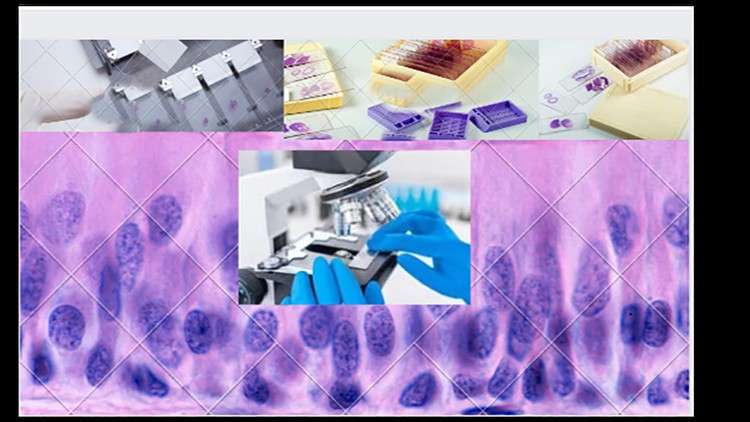
4 primary kinds of human tissue could be stained and considered utilizing numerous histological strategies. Epithelium, connective tissue, muscle tissue, and nervous tissue have commonalities however look very distinct structurally after staining. Every stain exists to focus on an essential characteristic or element inside a tissue kind. For instance, probably the most widespread stains, Hematoxylin, is a primary dye that stains proteins a blue colour, whereas Eosin stains proteins a pink colour. These two stains are generally used collectively to outline intracellular organelles and proteins. Due to the number of the proteins that exist, some stains had been created to focus on a selected protein, which this evaluation will talk about within the following sections. The good thing about utilizing a particular stain is that it will probably spotlight the particular protein very properly. Nonetheless, due to its specificity, the opposite constructions is not going to be seen. Because of this, a number of slides will typically be created from a given specimen in order that a number of stains could be carried out to collect the complete vary of wanted info.
Nearly all tissue stains are carried out on tissue that has been faraway from the physique. Nonetheless, in uncommon cases, very specialize stains known as very important stains can work on tissue remaining within the physique. These stains are used for the identification of particular kinds of tissue and identification of irregular tissue, so a subsequent biopsy could be extra correct in acquiring irregular tissue
Tissue Preparation
Earlier than particular staining can happen, tissue samples should bear preparation by means of the next levels: Fixation, processing, embedding, sectioning, and generally antigen retrieval. In fashionable histology laboratories, most of those steps are automated.
.Fixation: Fixation makes use of chemical compounds to protect the construction of the tissue in its pure kind and protects it from degradation by irreversibly cross-linking proteins. Though a number of specialised fixatives can be found, Impartial Buffered Formalin is a standard alternative for this step. The fixation step is significant to the remainder of the histologic staining process as a result of by retaining the chemical composition of the tissue, the pattern is hardened and makes the sectioning section simpler. Paraffin-formalin is one other efficient fixative. Its profit is that it’s the fixative of alternative for immunostaining; nevertheless, it requires preparation on the time of the fixation. Bouin is a fixative used for analyzing embryo and mind tissue due to its superior preservation of delicate nuclei and glycogen. Its draw back is that it doesn’t protect kidney tissues properly and in addition distorts mitochondrial construction.[1]
Dehydration: The addition of ethanol accomplishes the dehydration of a pattern. It eliminated water from the pattern and additional hardens the tissue for eventual mild microscopy. After ethanol is utilized, and following the completion of tissue dehydration, xylene is used to take away the ethanol.[1]
Embedding: Embedding is the method of placing the pattern right into a paraffin wax or a plastic resin to boost the method of extracting mobile constructions. This step is to be carried out with warning if the aim is to carry out immunostaining as a result of the paraffin wax will inhibit the penetration of antibodies, and result in a false outcome.[1]
Sectioning: Sectioning entails mounting the specimen on a microtome and reducing it into sections. The popular thickness is 4-5 micrometers in order that it may be stained and placed on a microscope slide for examination.[1]
Antigen Retrieval: This step is to retrieve antigens that would have been coated within the fixation and embedding levels. If the cross-linking of proteins conceals the antigen websites, there might not be as sturdy of an immunohistochemical response. Antigen retrieval is achieved by means of heating and proteolytic strategies to interrupt down the cross-links and reveal the epitopes and antigens that had been beforehand coated.[1] Though this step carries the danger of denaturing each the fixative and the antigens themselves, a profitable antigen retrieval technique can result in a way more efficient immunostaining depth.
Content material
Tissue – Epithelium
Histology blood
Bone histology
histology cartilage
Histology of respiratory system
histology of circulatory system
connective tissue

The put up histology fundamentals for medical college students appeared first on destinforeverything.com/cms.
Please Wait 10 Sec After Clicking the "Enroll For Free" button.